Neck Lymph Nodes

The neck can be divided into ‘central’ and ‘lateral’ compartments. The central compartment contains the thyroid gland, parathyroid glands, central neck lymph nodes neck, voice box and swallowing apparatus. The lateral compartment contains salivary glands, lymph nodes, blood vessels and nerves.

Lymph nodes are small bean-shaped structures that filter a fluid called ‘lymph’. Lymph is carried by lymphatics which are small channels within the neck. As lymph passes through lymph nodes, the immune cells contained within the nodes check the lymph for germs, cells or foreign substances. If anything abnormal is identified, the immune cells within the nodes are activated and mount an immune response to help rid the body of the foreign material.

When the cells within lymph nodes are active fighting infection or inflammation, the lymph node can grow and form a lump in the neck. This is the most common reason we see enlarged lymph nodes in the neck. Generally, once the infection or inflammation settles, the lymph node gradually decreases in size over weeks back to its baseline size.

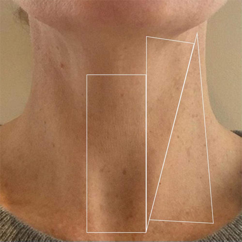
Central Compartment (Central Neck)

1 of 3

Anterior (Front) Compart-ment of Lateral Neck

2 of 3

Posterior (Back) Compartment of Lateral Neck

3 of 3

However, occasionally a lymph node can become large for other reasons. Cancer arising in the mouth, throat, voice box, skin, salivary glands or thyroid can spread to lymph nodes in the neck and cause lymph node enlargement. Similarly, cancer within the lymph nodes (called ‘lymphoma’) can also cause node enlargement. The location of the cancer often determines where in the neck the lymph nodes will first enlarge.

As a good rule of thumb, if you notice a lump in your neck that is enlarging or not resolving over a 4 week period, it should be looked at by your doctor. They will either order a neck ultrasound or refer you to a specialist who does their own ultrasounds. The ultrasound can determine whether a lymph node is enlarged and whether its appearance is normal or abnormal. These findings guide whether a biopsy (‘fine needle aspiration’ or ‘core biopsy’) is indicated. Details of this procedure can be found on this website under the ‘Ultrasound’ tab.

Ultrasound appearance of a normal “benign” lymph node

Ultrasound appearance of a pathologic, atypical lymph node

Lymph node surgery

Lymph nodes may require surgical removal for diagnostic purposes. For example, if the needle biopsy result is inconclusive, removal of a whole lymph node for microscopic examination may be necessary to secure a diagnosis. Such ‘open’ lymph node biopsies are done through a small neck incision made directly over the lymph node in question. 


Lymph nodes may also require surgical removal for treatment purposes. For example, if the lymph node is enlarged because of spread form a primary cancer in one of the locations mentioned previously, part of the treatment for that cancer may involve removing all lymph nodes draining that primary site of cancer. This surgery is done through an incision on the neck and great care is taken to preserve non lymphoid structures such as nerves and blood vessels. Dr Sinclair will discuss such a procedure in detail with you if this type of surgery is required as part of your treatment course.

Scroll to Top