Ultrasound is a non-invasive imaging technique that uses high-energy sound waves to look at tissues in the body. An ultrasound examination of the neck is an integral part of the work up of a neck, salivary or thyroid mass, or to look for enlarged parathyroid glands. The ultrasound is also essential to guide needle placement for biopsies of appropriate neck lesions and to guide electrode placement during radiofrequency ablation of thyroid nodules.
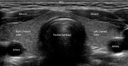
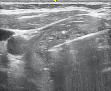
A/Prof Sinclair has performed over 12000 neck ultrasound examinations in the past decade and over 3000 neck and thyroid biopsies. She has published and lectured widely on the use of ultrasound for thyroid nodule assessment, thyroid cancer surveillance, and neck lymph node assessment. She sees the ultrasound as a ‘third arm’ in the work-up of neck lesions and finds it an indispensable tool to help individualise surgical and non-surgical approaches to thyroid nodules, thereby assuring preservation of as much normal thyroid tissue as possible . A normal thyroid ultrasound is shown.
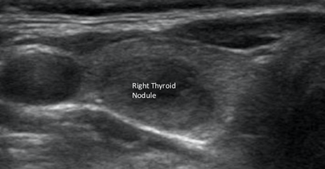
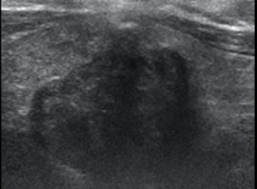
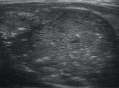
Thyroid Nodules on Ultrasound
Nodules appear as ‘lumps’ inside the thyroid gland.
The size and appearance of a given thyroid nodule are used to determine whether a biopsy is needed. Features of a nodule that increase the risk of cancer include solidity, hypoechoic appearance, calcification, irregular margins, taller than wide shape, and extension of the nodule outside of the thyroid gland (see images below).
Thyroid, Neck and Salivary Biopsies (Fine Needle Aspiration - FNA)
Fine needle aspiration is the process by which cells are removed from a thyroid, neck or salivary mass to allow for laboratory analysis and determination of cancer risk. This is generally a safe procedure provided the person performing the biopsy is skilled in ultrasound-guided neck biopsies.
During an FNA procedure, an injection of local anesthetic will be placed in the skin before the biopsy is performed. Once the skin is numb, a thin needle will be inserted through the skin and into the neck lesion of interest, under direct ultrasound visualization. Cells will be sampled and the needle removed. For any given neck lesion, A/Prof Sinclair usually performs three FNA’s which minimizes the chance of a ‘non-diagnostic’ biopsy result. Please contact Dr Sinclair for further information or questions related to neck ultrasound or FNA biopsies.